Keratoconus literally means cone-shaped cornea (AAO quote 2010)
The cornea is the window and outer surface of the eye. When you are interpreting an image light travels through the cornea past the lens to the retina and then the brain to form a visual image. The normal corneal surface is smooth and aspheric and flattens towards the edges. Light rays passing through it moves in an undistorted manner to the retina to project a clear image to the brain. This is the typical normal working cornea.
Keratoconus is a very slow progressive eye condition that affects the cornea. The normally round, dome-shaped cornea weakens and thins, causing a cone-like bulge to develop. The regular curvature of the cornea becomes irregular, resulting in increasing nearsightedness (myopia) and astigmatism that have to be corrected with special glasses or contact lenses. Since the cornea is responsible for refracting most of the light coming into the eye, corneal abnormalities can result in significant visual impairment, making simple tasks like driving or reading books.
(EYE FACTS AAO worksheet, 2010)
About 1/ 2000 people will develop keratoconus. Most people will have a mild or moderate form of the disease. Less than 10% of people with keratoconus will develop the most severe form. It typically is diagnosed in the late teens or twenties. However, many people have been diagnosed in their mid to late thirties; this is usually a more mild form of the disease. It is common for one eye to precede faster than the other and the eyes may go for long periods of time without any change and then change dramatically over a period of months.
The treatment protocol will depend on the level of keratoconus. Sometimes vision might be correctable with glasses or rigid contact lenses. When vision worsens and the preliminary treatments are not effective a cornea transplant might be necessary. According to the American Academy of Ophthalmology about 10-20% of keratoconus patients require a corneal transplant from a donor cornea. When this happens the ophthalmologist will essentially remove the diseased cornea and replace it with a healthy one.
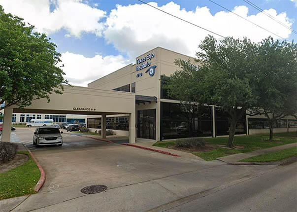
If you are a Texas keratoconus patient your eye doctor will need your cooperation. Since the doctors have no control over what happens in between visits you will need to remind yourself that these opthalmologists are your consultants. The eye doctors here at the Texas Eye Institute will be happy to diagnose keratoconus and suggest a proper treatment protocol. It is up to keratoconus patients to ultimately be proactive and care for their eyes. Also remember if you are seeing any other health professional, be sure they know about your keratoconus. Once again if you think you may have keratoconus and are seeking a houston keratoconus eye doctor please call us at the Texas Eye Institute.
Texas Eye Institute is proud to provide five convenient locations for your eye care needs. Visit one of our convenient locations in Angleton, Sugarland, Southwest Houston, Katy, or Southeast Houston to see why the Texas Eye Institute is the best choice to care for your vision. Need LASIK in Houston? What about a comprehensive eye exam in Sugarland? See our locations page to find our practice nearest you!


